Histopathology Images
Endometrial Stromal Sarcoma |
|
 |
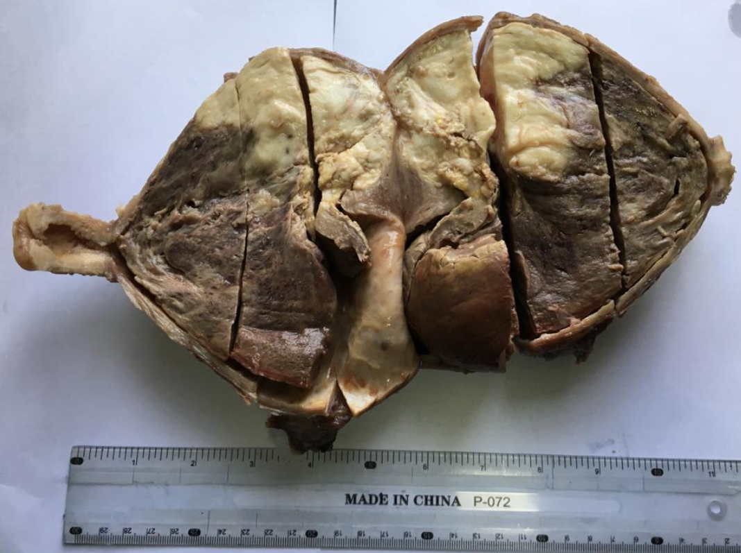 |
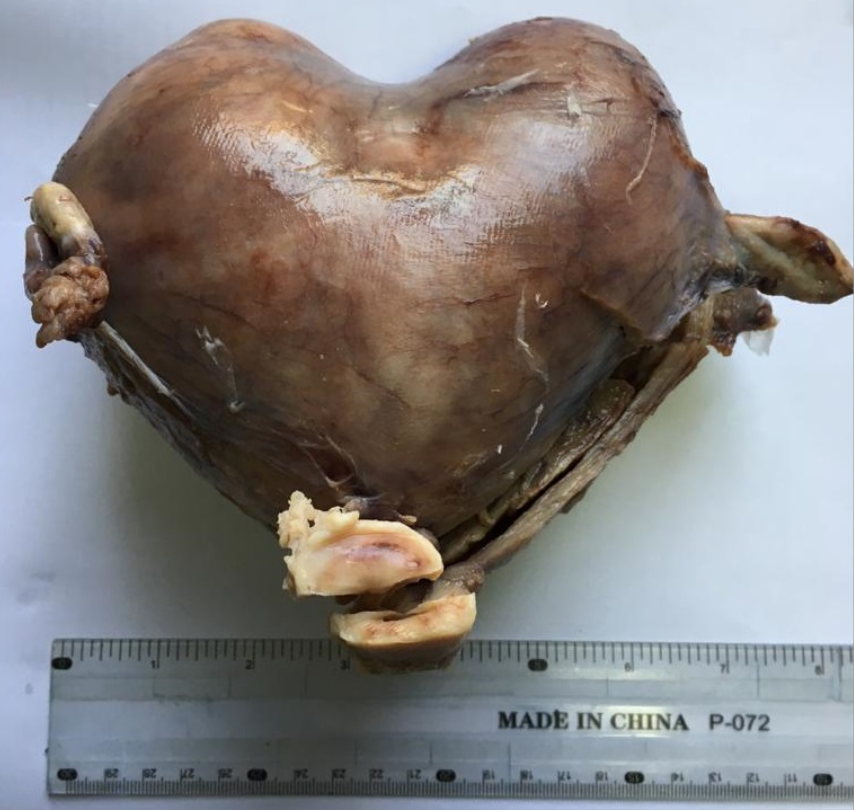
Figure 1: Gross presentation of endometrial stromal sarcoma. Intact bicornuate uterus appears heart-shaped. |
Figure 2: Endometrial stromal sarcoma. Cut surface of uterus shows large necrotic mass obliterating the endometrial cavity and reaching up to isthmus. |
 |
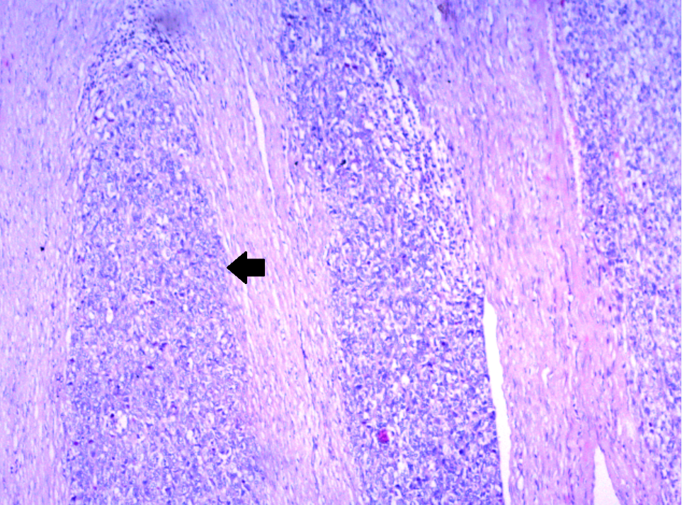 |
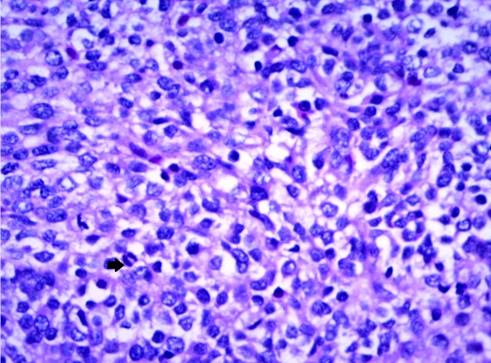
| Figure 3: Microscopic view of endometrial stromal sarcoma with pleomorphic nuclei and marked mitosis (arrow) (H&E, 400x). | Figure 4: Microscopic view of high grade endometrial stromal sarcoma infiltrating the myometrium (arrow) (H&E, 100x). |
| Metaplastic Carcinoma of Breast | |
 |
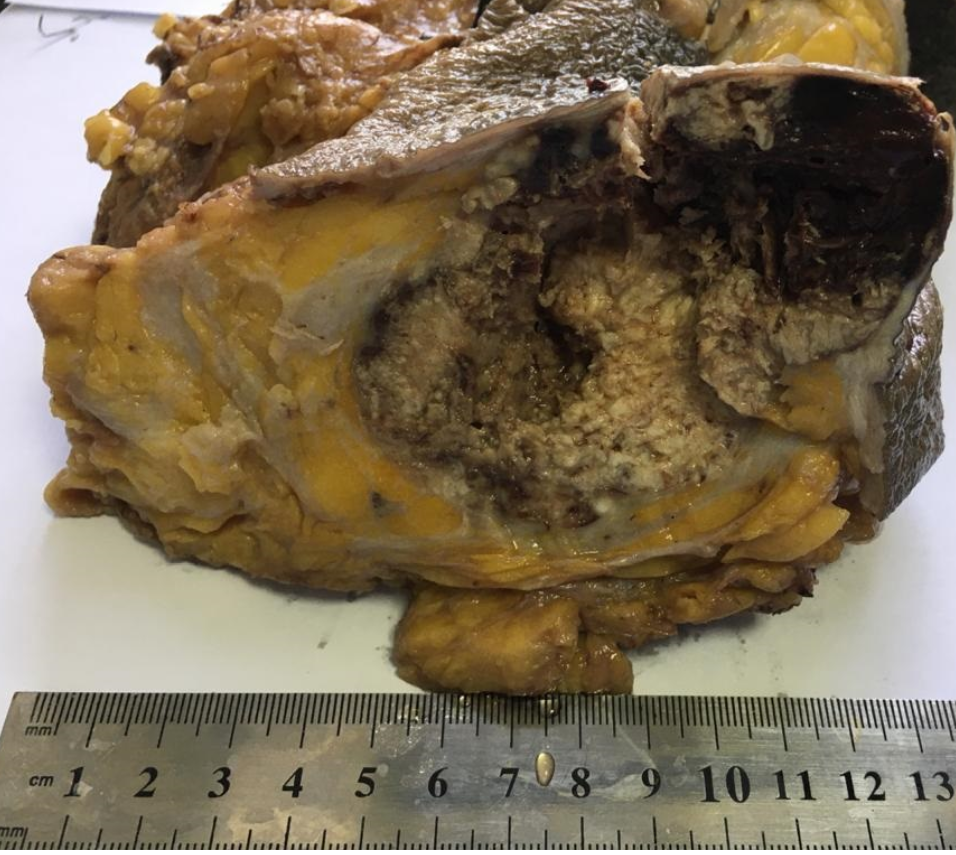 |
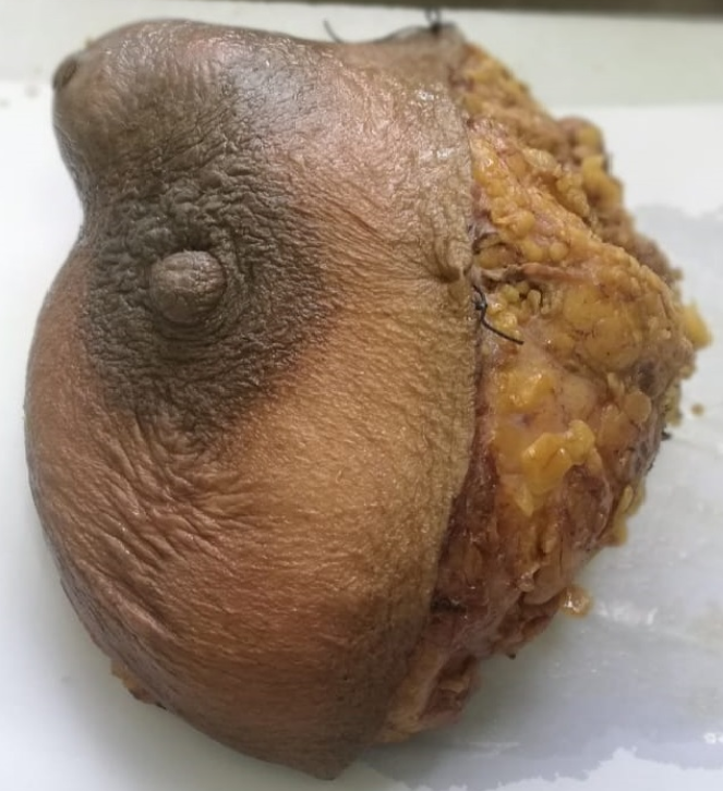
| Figure 1: Gross appearance of mastectomy specimen with large prominent lump. | Figure 2: Cut surface of mastectomy specimen showing large necrotic mass with hemorrhagic areas. |
 |
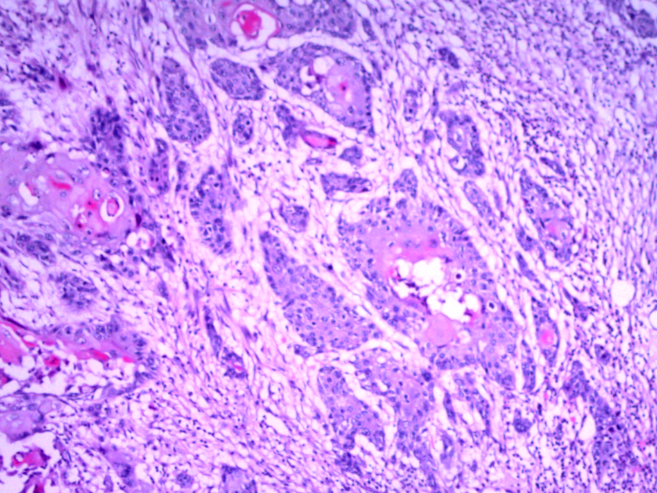 |
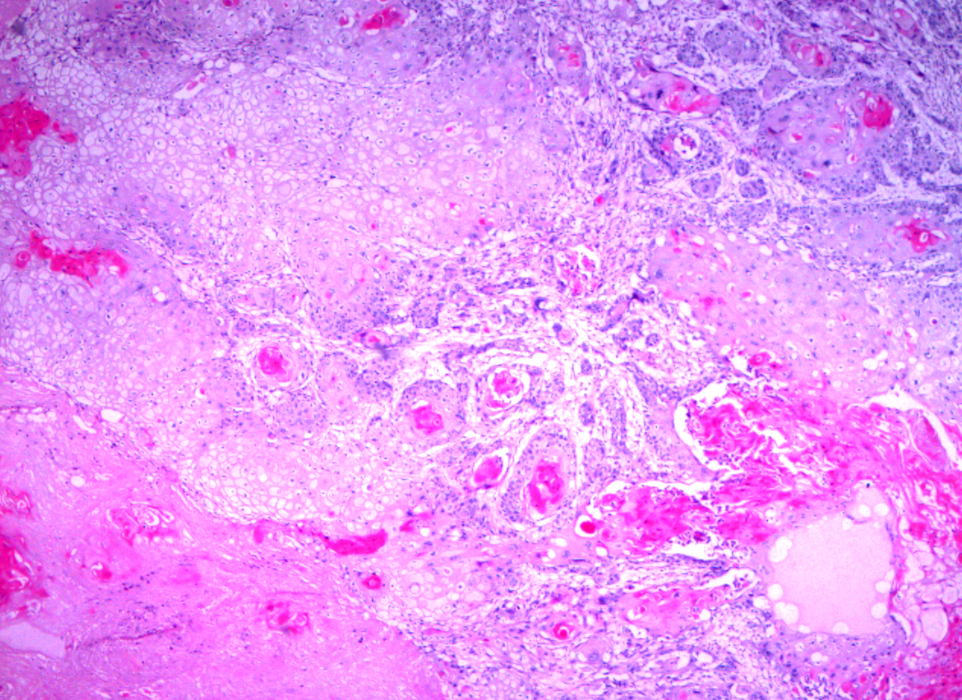
| Figure 3: Microscopic examination reveals pure squamous cell carcinoma (metaplastic carcinoma) of the breast (H&E, 100x). | Figure 4: Metaplastic carcinoma of breast showing nests of squamous cells infiltrating the stroma (H&E, 100x). |

Volume 9 Issue 4
An official publication of
Islamabad Medical and Dental College.
Contributed by:
Prof Dr. Saeed Alam, Dr. Javeria Faridi
Department of Pathology
Islamabad Medical & Dental College