Hypertonic Dextrose Prolotherapy for Knee Osteoarthritis: A Comprehensive Review of Mechanism, Techniques, Limitations, and Side Effects
Rao Erbaz Hassan1, Yaseen Ahmad2, Hamza Haroon2, Arif Ullah Khan1, Mahnoor Abbasi3
1Department of Trauma and Orthopedics, Khyber Teaching Hospital, Peshawar; 2Department of Trauma and Orthopedics, Lady Reading Hospital, Peshawar; 3Department of General Surgery, Ayub Teaching Hospital, Abbottabad.
This review delves into the potential of hypertonic dextrose prolotherapy (HDP) as a treatment for knee osteoarthritis (KOA), a prevalent condition, that significantly impacts individuals' quality of life, particularly affecting older adults. HDP involves injecting a hypertonic dextrose solution into or around the affected joint, inducing inflammation and subsequent tissue healing. This review examines various aspects of HDP, including its mechanism of action, comparative effectiveness with other therapies, dosage, and safety considerations as well as HDP's cost-effectiveness, safety profile, and efficacy in providing pain relief and improving function. Comparative studies indicate its superior effectiveness compared to other therapies such as saline, exercise, and steroids, with persistent benefits observed over extended follow-up periods. However, careful consideration of contraindications and potential side effects is crucial. Further research is warranted to conclusively establish its safety and long-term efficacy. In summary, HDP offers a promising avenue for managing KOA, potentially improving outcomes and enhancing the quality of life for affected individuals.
Keywords: hypertonic dextrose, peri-articular injections, intra-articular injections, intra-articular knee pathology, knee osteo-arthritis, kellgren–lawrence scale, persistent knee pain, prolotherapy, dextrose prolotherapy
Osteoarthritis (OA) is the most common form of arthritis, affecting about 15% of the world's population1. Knee OA (KOA) is the most frequently diagnosed joint site, with a lifetime risk of developing symptomatic KOA at 45%. Approximately 32.5 million adults in the U.S. have been clinically diagnosed with OA, and of these, 14 million have symptomatic KOA. About 6 million are between the ages of 45 and 64, and another 6 million are 65 or older. The prevalence of symptomatic KOA is higher in women across all age groups2. The progression of KOA is generally slow and can take years, with the disease often going through stages or gradually worsening over time3. KOA involves cartilage damage and is influenced by factors such as aging, genetics, metabolic syndrome, obesity, repetitive use of joints, bone density, muscle weakness, and trauma. Key factors in OA progression include the degradation of cartilage by enzymes like aggrecanase and collagenase, driven by inflammation and mechanical stress. The infrapatellar fat pad and synovial membrane form a functional unit that contributes to OA through inflammatory pathways. Understanding these pathways is crucial for guiding the treatment of KOA4. X-rays are the primary method for diagnosing KOA. The Kellgren-Lawrence scale, a grading system based on AP knee radiographs, categorizes OA severity from 0 (no OA) to 4 (severe OA with significant joint narrowing, marked sclerosis and bone deformity, and large osteophytes)5 (Figure 1). While most OA patients eventually need knee replacement, they can typically manage with pain medication and other non-surgical treatments for an average of 13 years before surgery is necessary2. Since 1950s, prolotherapy (Proliferation therapy) has been used as a treatment for knee osteoarthritis. Dr. Hackett's work in 1958 laid the groundwork for this approach6. While other substances have been explored, the most common and well-studied injection used in prolotherapy for KOA is a hypertonic dextrose solution7. When injected, the hypertonic dextrose induces mild cellular injury via a rapid osmotic shift of fluid, initiating an inflammatory response8. Prolotherapy offers a cost-effective treatment option for KOA. This review explores the mechanism, technique, limitations, and side effects of hypertonic dextrose prolotherapy (HDP) for KOA.
Review:
Knee osteoarthritis management includes various interventional techniques beyond traditional options like corticosteroid injections and viscosupplementation. Newer methods such as prolotherapy with hypertonic dextrose (HD), platelet-rich plasma (PRP), stem cells, and bone marrow aspirate are being extensively investigated for their effectiveness. Genicular nerve blocks with radiofrequency ablation or cryoneurolysis are also emerging as promising alternatives. Each method has a distinct mechanism of action and clinical impact. These techniques provide pain relief and may help patients delay or avoid knee replacement surgery9,10. HDP has gained attention of clinicians due to its cost-effectiveness, safety profile and efficacy.
HD is thought to be an ideal choice for prolotherapy because it dissolves easily in water, is a natural component of blood, and can be safely injected in large amounts into various tissues. For decades, clinicians have used HD injections, both intra-articularly and extra-articularly, to treat musculoskeletal pain. Recent years have seen an increase in controlled trials investigating its effectiveness in treating symptomatic KOA10.
Mechanism of action:
HDP likely work through a combination of factors. The high sugar concentration dehydrates cells at the injection site, causing some to lyse and triggering an initial inflammatory response11,12. This inflammation is followed by a healing process driven by cells like granulocytes and macrophages. The initial inflammation and the later healing phase are interconnected. This controlled stress response seems to activate the inherent healing ability of injured soft tissues12,13,14. Inflammatory cytokines like Prostaglandins, along with other factors, regulate the cellular environment that guides tissue repair.
Cells from the initial inflammation stage (granulocytes and macrophages) may release signals attracting and activating fibroblasts, which are key players in collagen production. This collagen deposition helps rebuild the tissue until stability is restored. The newly formed collagen matures and tightens, potentially strengthening the tendon or ligament.12 Reeves et al., discussed also cartilage proliferation mechanisms based on human studies 15. There is no compelling evidence to confirm or deny cartilage proliferation, although a small sample study supports its occurrence15,16
Larger studies with more effective designs are needed to establish this fact.
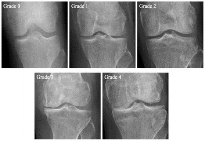
Figure 1: The example of the Kellgren–Lawrence scale. In grade 0, there are no pathological features of KOA. Grade 1 shows potential joint space narrowing with doubtful osteophyte formation. Grade 2 demonstrates definite osteophyte formation and possible
Technique: Intra-articular versus peri-articular
The site of injection has been a topic of interest in recent times. 17-19 Supra lateral 19,20 and inferiolateral 17 are the most common techniques used for intra-articular method. Risk of intra-articular infection and hemearthrosis are possible side effects to this technique.18 For peri-articular injection, different techniques have been used with comparable efficacy observed.17,18 Multiple points are injected in different sites of knee for this purpose (Figure 2). Pain during these multiple infections and post-injection pain are the usual points of concern for the patients18. From Rezasoltani et al. (2017).18 In terms of results, both techniques are comparable.17,18 While the peri-articular technique may provide more pain relief, shorter pain periods, and less joint limitation, both techniques improve morning stiffness, difficulty rising from a seated position, joint locking, walking, climbing stairs, and sitting/standing pain.18 The better pain alleviation profile of peri-articular injections can be attributed to the lower concentration of HD used, as its analgesic ability at lower doses (5-15%) has been documented.12
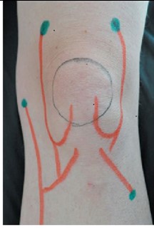
Figure 2: Points of peri-articular injection around the knee are indicated by blue dots, while the articular nerves of the left knee are shown as red lines.
In order to achieve the optimal benefits of HDP, different researchers have used both intra-articular and peri-articular techniques simultaneously21,22. This approach seems plausible as it avoids any unwanted bias and provides optimal relief.
Comparison with other therapies
Cortez et al. 23 compared HDP to other treatments for knee osteoarthritis (KOA) using WOMAC and VAS scores. They found HDP superior to saline, physical therapy, and exercise, but inferior to PRP, erythropoietin, and autologous conditioned serum. HDP was similar to radiofrequency, botulinum toxin A, and hyaluronic acid in some studies. Another review by Wee et al.24 also yielded mixed results, but suggested potential benefits of HDP. However, this review raised concerns about bias in the studies. Notably, HDP showed similar pain relief to PRP but was less effective in reducing stiffness at 6 months.24 No studies have yet compared the effectiveness of HDP injections to corticosteroids for treating knee osteoarthritis.24 This presents a valuable research opportunity. It would be interesting to see if locally injected HD offers similar benefits in knee osteoarthritis as it has shown promise in treating other musculoskeletal conditions, such as general musculoskeletal pain25, supraspinatus tendinopathy,26 and carpal tunnel syndrome27. HDP's effectiveness in treating chronic musculoskeletal pain has been compared to saline, exercise, PRP, and steroids in several studies. A systematic review revealed that HDP significantly reduced pain scores compared to saline at 6 months to 1 year of therapy, but not at other time points. Compared to exercise, HDP showed greater pain reduction from 1 to 3 months and 6 to 12 months, but no difference was observed at baseline.25
When compared to PRP, HDP showed similar effectiveness with no significant differences in pain reduction. Similarly, HDP was comparable to steroids in terms of therapeutic effects.25 HDP is a relatively new and safe treatment option for KOA. It may be a good choice for patients who cannot afford other options, have exhausted other therapies, are poor surgical candidates, or are not willing to go for surgery.
Dosage and duration of therapeutic effect:
According to available literature, HDP has been used in grade 2 and 3 KOA.13, 17-19 The number of doses ranges from 1 to 5, with 3 being the most common, and intervals vary from weekly to every two months, typically monthly. Intra-articular injections generally use a 25% dextrose concentration with a volume of 2-8 ml, while peri-articular injections typically use a 15% dextrose concentration. 2-6 sessions at monthly intervals may be ideal.25,28 Multiple studies on HDP effects in treating KOA with follow-up periods of 2-3 months, 5-6 months, and 12 months show persistent benefits.28 Rabago et al. reported these beneficial effects lasting up to 2.5 years.29 These findings suggest that HDP offers long-term advantages, potentially outlasting other treatments like steroids and hyaluronic acid, and comparable to PRP.28
Contraindications
HDP for KOA has few absolute contraindications, similar to other injectable treatments. Active infections near the injection site (cellulitis) or within the joint (septic arthritis) are absolute contraindications due to the risk of spreading infection. Additionally, acute inflammatory conditions such as acute gouty arthritis and acute flares of rheumatoid arthritis can worsen with HDP due to their inflammatory nature. Acute fractures require proper healing, and prolotherapy might disrupt this process. Although uncommon, an allergy to corn is a contraindication for HDP.28,30 Medical professionals should carefully assess patients for these contraindications before proceeding with prolotherapy for KOA.
Common side effects and adverse effects
While studies haven't reported adverse events in HDP for KOA, potential safety concerns remain. Injecting irritant solutions near tendons, ligaments, and joints carries inherent risks.
Existing studies lack sufficient power to detect uncommon adverse events. Potential complications include infection, allergic reactions to dextrose, lightheadedness, and nerve damage. Recommendations for safe practice include adhering to universal precautions to minimize infection risk, positioning patients prone or supine to reduce vasovagal episodes, and monitoring for potential side effects, including mild to moderate pain, inflammation, and self-limiting hematomas.28,30
HDP for KOA appears to have a low-risk profile, but further studies are needed to definitively assess its safety, particularly regarding uncommon adverse events. HDP has gained interest for its potential benefits: affordability, safety, and effectiveness. It's believed to work by inducing inflammation through dehydration, which then triggers a healing process involving collagen deposition. While studies since its introduction in 1958 have been promising, high-quality, large-scale research is lacking. Conducting such studies would be crucial for establishing proper guidelines for HDP use.
In conclusion, HDP presents a promising avenue for managing KOA. Its cost-effectiveness, safety profile, and demonstrated efficacy in providing pain relief and improving function position it as a viable treatment option. Comparative studies have shown its superior effectiveness compared to other therapies such as saline, exercise, and steroids, with persistent benefits observed over extended follow-up periods. However, careful consideration of contraindications and potential side effects is essential, and further research is needed to conclusively ascertain its safety and long-term efficacy. Overall, HDP represents a novel and promising addition to the multifaceted approach to managing KOA, offering hope for improved outcomes and enhanced quality of life for affected individuals.
- Callahan LF, Cleveland RJ, Allen KD, Golightly Y: Racial/ethnic, socioeconomic, and geographic disparities in the epidemiology of knee and hip osteoarthritis. Rheum Dis Clin North Am. 2021, 47:1-20. https://doi.org/10.1016/j.rdc.2020.09.001
- Arthritis by the numbers. (2020). Accessed: March 3, 2024: https://www.arthritis.org/getmedia/73a9f02d-7f91-4084-91c3-0ed0b11c5814/abtn-2020-final.pdf.
- Martel-Pelletier J, Barr AJ, Cicuttini FM, et al.: Osteoarthritis. Nat. Rev. Dis. Primers. 2016, 2:16072. https://doi.org/10.1038/nrdp.2016.72
- Jang S, Lee K, Ju JH: Recent Updates of Diagnosis, Pathophysiology, and Treatment on Osteoarthritis of the Knee. Int. J. Mol. Sci. 2021, 22:2619. https://doi.org/10.3390/ijms22052619
- Kohn MD, Sassoon AA, Fernando ND: Classifications in Brief: Kellgren-Lawrence Classification of Osteoarthritis. Clin Orthop Relat Res. 2016, 474:1886-93. https://doi.org/10.1007/s11999-016-4732-4
- Hackett GS: Ligament and Tendon Relaxation (Skeletal Disability) treated by Prolotherapy (Fibro-Osseous Proliferation). 3rd ed. Charles C Thomas, Springfield, Illinois; 1958.
- DeChellis DM, Cortazzo MH: Regenerative medicine in the field of pain medicine: Prolotherapy, platelet- rich plasma, and stem cell therapy—theory and evidence. Tech Reg Anesth Pain Manag. 2011, 15:74-80. https://doi.org/10.1053/j.trap.2011.05.002
- Jensen KT, Rabago DP, Best TM, Patterson JJ, Vanderby R Jr: Early inflammatory response of knee ligaments to prolotherapy in a rat model. J Orthop Res. 2008, 26:816-23. https://doi.org/10.1002/jor.20600
- Malik KN, Camp N, Chan J, Ballard M: Interventional techniques for the management of knee osteoarthritis: A literature review. Cureus. 2023, 15: e47133. 10.7759/cureus.47133
- Wang J, Liang J, Yao J, et al.: Meta-analysis of clinical trials focusing on hypertonic dextrose prolotherapy (HDP) for knee osteoarthritis. Aging Clin Exp Res. 2022, 34:715-24. https://doi.org/10.1007/s40520-021-01963-3
- Linetsky FS, FRafael M, Saberski L: Pain management with regenerative injection therapy (RIT) Weiner RS, ed. Pain Management. Pain Management 6th ed. Weiner RS (ed): CRC Press, Boca Raton, FL; 2002. 381-402. https://doi.org/10.1201/9781420093193
- Wilson M, Topping A, Praet S: Dextrose Prolotherapy - Mechanism of Action. Act Scie Ortho. 2022, 149-56. https://doi.org/10.31080/asor.2022.05.0585
- Reeves KD, Hassanein K: Randomized prospective double-blind placebo-controlled study of dextrose prolotherapy for knee osteoarthritis with or without ACL laxity. Altern Ther Health Med. 2000, 6:68-80.
- Topol GA, Reeves KD, Hassanein KM: Efficacy of dextrose prolotherapy in elite male kicking-sport athletes with chronic groin pain. Arch Phys Med Rehabil. 2005, 86:697-702. https://doi.org/10.1016/j.apmr.2004.10.007
- Reeves KD, Sit RWS, Rabago DP: Dextrose prolotherapy. Phys Med Rehabil Clin N Am. 2016, 27:783-823. https://doi.org/10.1016/j.pmr.2016.06.001
- Topol GA, Podesta LA, Reeves KD, et al.: The chondrogenic effect of intraarticular hypertonic-dextrose (prolotherapy) in severe knee osteoarthritis. PM R. 2016, 8:1072-82. https://doi.org/10.1016/j.pmrj.2016.03.008
- Farpour HR, Fereydooni F: Comparative effectiveness of intra-articular prolotherapy versus peri-articular prolotherapy on pain reduction and improving function in patients with knee osteoarthritis: A randomized clinical trial. Electron Physician. 9(11): 5663-5669. 10.19082/5663. 2017, 9:5663-9. https://doi.org/10.19082/5663
- Rezasoltani Z, Taheri M, Kazempour Mofrad M, Mohajerani S: Periarticular dextrose prolotherapy instead of intra-articular injection for pain and functional improvement in knee osteoarthritis. J Pain Res. 2017, 10:1179-87. 10.2147/jpr. s127633
- Sit RWS, Wu RWK, Rabago D, et al.: Efficacy of intra-articular hypertonic dextrose (prolotherapy) for knee osteoarthritis: A randomized controlled trial. Ann Fam Med. 2020, 18:235-42. https://doi.org/10.1370/afm.2520
- Sert AT, Sen EI, Esmaeilzadeh S, Ozcan E: The effects of dextrose prolotherapy in symptomatic knee osteoarthritis: A randomized controlled study. J Altern Complement Med. 2020, 26:409-17. https://doi.org/10.1089/acm.2019.0335
- Rabago D, Patterson JJ, Mundt M, et al.: Dextrose prolotherapy for knee osteoarthritis: a randomized controlled trial. Ann Fam Med. 2013, 11:229-37. https://doi.org/10.1370/afm.1504
- Eroglu A, Sari A, Durmus B: Platelet-rich plasma vs prolotherapy in the management of knee osteoarthritis: Randomized placebo-controlled trial. Turk J Sports Med. 2017, 51:34-43. https://doi.org/10.5152/tjsm.2016.005
- Cortez VS, Moraes WA, Taba JV, et al. Comparing dextrose prolotherapy with other substances in knee osteoarthritis pain relief: A systematic review. Clinics. 2022, 77:100037.
- Wee TC, Neo EJR, Tan YL: Dextrose prolotherapy in knee osteoarthritis: A systematic review and meta-analysis. J Clin Orthop Trauma. 2021, 19:108-17.
- Bae G, Kim S, Lee S, Lee WY, Lim Y: Prolotherapy for the patients with chronic musculoskeletal pain: systematic review and meta-analysis. Anesth Pain Med. 2021, 16:81-95. https://doi.org/10.17085/apm.20078
- Cole B, Lam P, Hackett L, Murrell GAC: Ultrasound-guided injections for supraspinatus tendinopathy: corticosteroid versus glucose prolotherapy - a randomized controlled clinical trial. Shoulder Elbow. 2018, 10:170-8.
- Babaei-Ghazani A, Moradnia S, Azar M, et al.: Ultrasound-guided 5% dextrose prolotherapy versus corticosteroid injection in carpal tunnel syndrome: A randomized, controlled clinical trial. Pain Manag. 2022, 12:687-97. https://doi.org/10.2217/pmt-2022-0018
- Arias-Vázquez PI, Tovilla-Zárate CA, Legorreta-Ramírez BG, et al.: Prolotherapy for knee osteoarthritis using hypertonic dextrose vs other interventional treatments: systematic review of clinical trials. Adv Rheumatol. 2019, 59:39. https://doi.org/10.1186/s42358-019-0083-7
- Rabago D, Mundt M, Zgierska A, Grettie J: Hypertonic dextrose injection (prolotherapy) for knee osteoarthritis: Long term outcomes. Complement Ther Med. 2015, 23:388-95. https://doi.org/10.1016/j.ctim.2015.04.003
- Nourani B, Rabago D: Prolotherapy for knee osteoarthritis: A descriptive review. Curr Phys Med Rehabil Rep. 2016, 4:42-9. https://doi.org/10.1007/s40141-016-0111-z

An Official Publication of
Islamabad Medical & Dental College
Volume 13 Issue 2
Review Article
Address of Correspondence
Hamza Haroon
Email:
hamzamashwani09@gmail.com
Authors’ Contribution: All authors contributed equally to the conception, literature search, manuscript drafting, editing and review
Cite this article. Hassan RA, Ahmad Y, Haroon H, Khan AU, Abbasi M. Hypertonic Dextrose Prolotherapy for Knee Osteoarthritis: A Comprehensive Review of Mechanism, Techniques, Limitations, and Side Effects. J Islamabad Med Dental Coll. 2024; 13(2): 375-380.
DOI: https://doi.org/10.35787/jimdc.v13i2.1245