Role of MDCT in Patients with Abdominal Trauma
Abdul Qadeer Khan1, Shaista Nayyar1, Tahira Hameed2, Adeel Azhar3, Fawad Yasin1, Nayab Khan1
1Department of Radiology, Pakistan Air Force Hospital (PAF), Islamabad, 2Department of Surgery, King Fahad Specialist Hospital, Buraydha, KSA, 3Department of Radiology, Combined Military Hospital (CMH), Kohat
Objective: To assess the role of abdominal imaging in trauma patients for precise identification of injuries by using multidetector computed tomography (MDCT) aiding in either conservative management or early surgical exploration of the patient.
Methodology: The study was carried out on 120 cases of abdominal trauma. MDCT scan with contrast was performed after the initial resuscitation and stability of the patients in the emergency room being done by the respective medical and paramedical staff.
Results: In clinically stable patients, the use of multi-detector CT as initial triage of abdominal trauma resulted in a reduction in the number of laparotomies by 91% and an increase in number of conservatively managed with a better outcome of around 95% and 92% respectively.
Conclusion: The current study assessed and showed the better role of MDCT in evaluation of the blunt trauma patients in early detection and timely surgical exploration. MDCT imaging test helps the clinicians in making an early and right choice of intervention. The high-resolution MPR, MIP, and 3-D volume reconstruction images assisted in displaying complex injuries, increasing diagnostic capacity, accuracy, associated vascular and bony pathologies, and reduced reporting time.
Keywords: Blunt Abdominal Trauma, Multi-slice Detector Computed Tomography.
Abdominal trauma is categorized into two types, blunt abdominal trauma (BAT) and penetrating abdominal trauma. 1 The rate of blunt abdominal trauma is very high around 80% arriving in an emergency. 2 They are involved in high morbidity and mortality. Furthermore, the patient with blunt trauma faces many other pathophysiologic issues like rupture of hollow viscus due to external pressure.3 Also, the abdominal hollow viscera might collapse along with lacerations.4 Therefore, early detection of organ hemorrhage is vital in patients of abdominal trauma as active bleeding remains one of the principal causes of mortality.5 It is reported that physical examination might miss around 45% of such injuries, making it an unreliable approach.6 Whereas, abdominal imaging plays an important role in precisely identifying the injuries for early surgical exploration and assessing those that can be managed conservatively.7 In hemodynamically stable patients, CT is usually a preferred choice.8 imaging tests like FAST or MDCT are required to identify the severity and types of organ injuries so that a reliable treatment plan can be created, either conservative or requiring surgical intervention.9 Earlier detection can be carried out by FAST scan which is time-saving and cost effective and identify the presence of free fluid in abdominopelvic regions however ultrasound has a low sensitivity of about 46%.10 An advancement in medical diagnostics is the MDCT scan with decreased scan time, better resolution with thin slices and reduced artefact level.11 MDCT also possesses a usual edge over other imaging tests as its scans can be generated keeping in line with the 5 grades of the American Association for the Surgery of Trauma (AAST).12 The aim of the current study is to assess the role of MDCT in evaluation of the blunt trauma patients for early detection and timely surgical exploration and compare it with those who can be managed conservatively.13
The cross-sectional study was conducted at Pakistan Air Force (PAF) Hospital, Islamabad, Pakistan, from November 2022 to November 2023 after approval from the ethical committee. The sample size was calculated using the WHO sample size calculator, keeping the anticipated population proportion (P) of PV at 0.05. The data was collected from 120 cases of abdominal trauma through non-probability convenient sampling. Scanning was performed at our setup utilizing 128 slice MDCT scanner by Philips and Medrad power pressure injector for contrast agent. Patients selected initially went through FAST scan for correlation with it.
Inclusion Criteria: Patients of trauma with clinical suspicion of intra-abdominal injury, hemodynamically stable patient and multi-trauma patient.
Exclusion criteria: Hemodynamically unstable patients and patients with free fluid in abdominopelvic cavities on FAST scan or obvious peritoneal signs and progressive abdominal distention were sent for surgery excluding them from the study. Pediatric patients weren’t included in the study. The injury severity score (ISS) was used as a standard to assess the severity of traumatic injury. The median was found to be 22.5 having a 13-43 range and an interquartile range of 19-25. At the start of the study, demographic profile, history, physical examination findings, time since injury and initial sonographic findings of the patients were recorded. Later, any change in the management plan after MDCT examination was recorded and compared with the patient clinical outcome at the end of the study. Patients were informed in detail of the risks and benefits of the study and the possible effects of the contrast and the availability of the emergency medicines on side if side effects happen. Then patients’ consent was taken and the CECT abdomen was performed on a 128-detector. The adjusted dose of non-iodinated contrast according to the age and weight was administered using power injector at an average rate of 3.25 mL/s. If Foleys catheter passed to the patient, it was clamped. Non-contrast CT scanning was performed with a window from the level of the diaphragm to ischial tuberosities using 10 mm thick sections and reconstruction at 1.25 mm intervals. Non-ionic, iodinated contrast was administered using an automatic dual-pressure injector as a bolus at rate of 2-3 ml/sec. Delayed imaging after 3-5 minutes was performed in suspected renal and urinary tract injuries. Multiplanar reformation (MPR), maximum intensity projection (MIP) and 3D reconstruction were done in every case using CT grading systems. The plan of management was compared prior to and after the scan. Free fluid was quantified using Federle and Jeffery system (1994). Data was entered in a Microsoft Excel sheet and was statistically analyzed using tables and charts like pie and bar charts for p-values by means of Fishers exact test, Chi-square test and McNemar tests. Using 2-sample t tests, continuous variables were tested which included age and ISS. Whereas, in chi-square tests, categorical variables were tested which included injured organ, gender and of mode of injury. The P value ≤ 0.05 was considered statistically significant, with a confidence interval of 95%.
The sample characteristics concerning gender distribution are given below (figure 1).
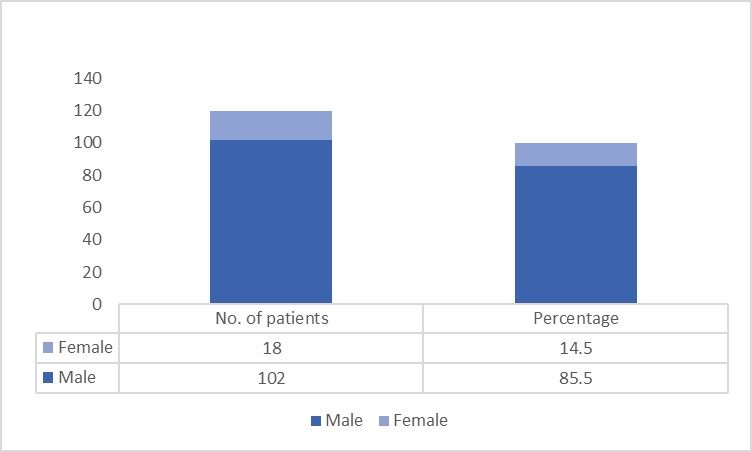
Figure 1: Gender Distribution of Patients
The participants belonged to different age groups, having a mean age of 21.40 ± 8.39 years. Our study found that the incidence of blunt abdominal trauma was relatively higher in youth in the age group of 20-30 years with a percentage of frequency of around 39% (figure 2).

Figure 2: Age Distribution of Patients
Our study showed that road traffic accidents (RTAs) was the most common cause of abdominal trauma. Of the 120 cases 82 (68%) patient’s had RTA, with the fall from height as the second most common with 22 (18.3%) frequency. Fall of heavy objects, sports injury, assaults and miscellaneous covered the rest (table I). Liver was the most commonly injured organ constituting 50 (41.6%) out of 120 cases followed by splenic injury 31 (25.8%). It was followed by injury of the kidney, urinary bladder and pancreas. Only 6 (5%) cases accounted for multiorgan injury while the rest of the 115 (95%) cases involved a single organ. Patients with multiple organs injured carried a high risk of mortality (table II). Liver hemorrhage (55%) was the most frequently observed hepatic injury followed by liver laceration (45%) (figure 3).
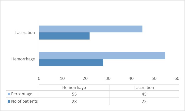
Figure 3: Liver Injury

Figure 4: Splenic Injury
In our study through better early diagnosis by MDCT, 109 (91%) injuries were managed conservatively whereas 11 (9%) injuries were managed surgically. Furthermore, the rate of negative MDCT in omitting injuries of an intra-abdominal nature was as low as 0.05%.
Table-I: Mode of Injury with Frequency |
||||||||
|
Mode of injury |
|
|
|||||
Frequency
|
|
Road traffic accidents |
Fall from height |
Fall of heavy objects |
Sports injury |
Assault |
Misc. |
Total |
No. of patients |
82 |
22 |
1 |
1 |
10 |
4 |
120 |
|
Percentage |
68 |
18.3 |
0.83 |
0.83 |
8.3 |
3.3 |
100 |
|
Age |
Mean |
42.87 |
45.54 |
55 |
33 |
45.2 |
50.5 |
43.83 |
SD |
±13.96 |
±15.70 |
0 |
0 |
±10.43 |
±18.57 |
±14.08 |
|
Table-II: Organ Involvement |
||||||||
|
Organ involvement |
|
||||||
Frequency |
|
Liver |
Spleen |
Kidney |
Urinary bladder |
Pancreas |
Multi-organ injury |
Total |
No. of patients |
50 |
31 |
21 |
2 |
10 |
6 |
120 |
|
Percentage |
41.6 |
25.8 |
17.5 |
1.6 |
8.3 |
5.0 |
100 |
|
Age |
Mean |
44.24 |
44.35 |
42.42 |
38 |
43.80 |
44.6 |
43.83 |
SD |
±13.92 |
±14.39 |
±15.67 |
±1.40 |
±12.37 |
±17.20 |
±14.08 |
|
In our study, we evaluated the role of MDCT in accurate and early detection of the abdominal visceral injuries.14 Although, the Focused Assessment with Sonography for Trauma (FAST) provides quick results, FAST imaging is not able to provide reliable results as per the surgical grades. Whereas, the MDCT provides the clinicians with options of treatment while considering the AAST scores.15 In our study, the most commonly injured organ was the liver. It was followed by injury of spleen, kidney, urinary bladder and pancreas. The results were similar to another study in which the hepatic injury was most common accounting for 34.78% (16/46) of cases.16 However, through the literature review we also found a contrasting result in regard to the incidence of organ damage. In a study conducted by Ibraheem et al. the most affected organ is Spleen (54%).17 Patients with multiple organs injured carried a high risk of mortality. In our study, only 6 (5%) cases accounted for multiorgan injury while rest of the 115 (95%) cases involved single organ. However, we were unable to find convincing literature evidence in this regard for the discussion. In a study, Road traffic accident (RTA) was found out to be the most common mode of injury accounting for 69.5% (32/46) of cases.18 Similarly, our study also showed that road traffic accidents (RTAs) were the most common cause of abdominal trauma. Of the 120 cases 82 (68%) patient’s had RTA. In our study through better early diagnosis by MDCT, 109 (91%) injuries were managed conservatively whereas 11 (9%) injuries were managed surgically. Similarly, in a study by Misra et al. conservatively managed cases were 60.86% of cases with the rest through surgical intervention.19 In our study there was increased accuracy through 3-D volume reconstruction images, high resolution MPR, and MIP. In case of hemoperitoneum identification and probability of visceral organ injury, MDCT is a reliable.
In our study, the rate of negative MDCT in omitting injuries of an intra-abdominal nature was as low as 0.05%. Similarly, a systematic review by Akeel et al. this rate is also reported as less than 0.06%. In another study, a significant lowering of laparotomies was observed due to the involvement of MDCT. The cases with conservative management also saw a significant rise, with decent result of around 95% and 92% respectively. Limiting factors include a small sample size and a retrospective single-institution study design. Further studies can be conducted at multiple centres to further strengthen our results.
The study concludes that MDCT imaging test helps the clinicians in making an early and right choice of intervention. MDCT-grade of solid organ injuries helps to treat patients through conservative management thus the need of surgical treatment will not be required. The high-resolution MPR, MIP, and 3-D volume reconstruction images assisted in displaying complex injuries, increasing diagnostic capacity, accuracy, associated vascular and bony pathologies, and reduced reporting time.
- Leppäniemi A. Nonoperative management of solid abdominal organ injuries: From past to present. Scandinavian Journal of Surgery. 2019 Jun;108(2):95-100. https://doi.org/10.1177/1457496919833220
- Caraiani C, Yi D, Petresc B, Dietrich C. Indications for abdominal imaging: When and what to choose?. Journal of ultrasonography. 2020 Apr 1;20(80):43-54. https://doi.org/10.15557/jou.2020.0008
- Nayak SR, Yeola MP, Nayak SR, Kamath K, Raghuwanshi PS. Role of focused assessment with sonography for trauma in the assessment of blunt abdominal trauma–a review. Journal of Evolution of Medical and Dental Sciences. 2021 Jan 4;10(1):45-51. https://doi.org/10.14260/jemds/2021/9
- Engles S, Saini NS, Rathore S. Emergency focused assessment with sonography in blunt trauma abdomen. International Journal of Applied and Basic Medical Research. 2019 Oct;9(4):193. https://doi.org/10.4103/ijabmr.IJABMR_273_19
- Boutros SM, Nassef MA, Abdel-Ghany AF. Blunt abdominal trauma: The role of focused abdominal sonography in assessment of organ injury and reducing the need for CT. Alexandria journal of Medicine. 2016 Mar 1;52(1):35-41. https://doi.org/10.1016/j.ajme.2015.02.001
- Gamberini L, Tartaglione M, Giugni A, Alban L, Allegri D, Coniglio C, Lupi C, Chiarini V, Mazzoli CA, Heusch-Lazzeri E, Tugnoli G. The role of prehospital ultrasound in reducing time to definitive care in abdominal trauma patients with moderate to severe liver and spleen injuries. Injury. 2022 May 1;53(5):1587-95. https://doi.org/10.1016/j.injury.2021.12.008
- Achatz G, Schwabe K, Brill S, Zischek C, Schmidt R, Friemert B, Beltzer C. Diagnostic options for blunt abdominal trauma. European Journal of Trauma and Emergency Surgery. 2020:1-5. https://doi.org/10.1007/s00068-020-01405-1
- Sultanoğlu H, Başaran A, Hekimoğlu A, Arslan ED, Özkan S. Ultrasound versus computed tomography scan findings in pediatric blunt abdominal traumas. 2022;6(3):e31 https://doi.org/10.18502/fem.v6i3.9393
- Madhu P, Santhosh D. Evaluation of Patients with Blunt Abdominal Injury by Ultrasonography (USG) and Multi Detector Computed Tomography (MDCT). International Journal of Contemporary Medicine Surgery and Radiology. 2019;4(4):D177-80. http://dx.doi.org/10.21276/ijcmsr.2019.4.4.42
- Tambe J, Mbuagbaw L, Nguefack-Tsague G, Foyet J, Ongolo-Zogo P. Multidetector computed tomography utilization in an urban sub-Saharan Africa setting: user characteristics, indications and appropriateness. The Pan African Medical Journal. 2020;37. https://doi.org/10.11604/pamj.2020.37.42.21176
- GOEL R, MAKHAIK S, JHOBTA A, SHAMI S, MAHAJAN P, KAPILA S. Role of MDCT in the Evaluation of Blunt Abdominal Trauma in Himalayan Region of Northern India. Journal of Clinical & Diagnostic Research. 2021 Nov 1;15(11)
- Kadeem MK, Abdulretha M. The Accuracy of Computed Tomography in Blunt Abdominal Trauma in unusual times. Prof.(Dr) RK Sharma. 2020 Oct;20(4):41819.
- Maqsood S, Khan TA, Ashraf S. Role of MDCT in blunt trauma abdomen. IAIM. 2018 Mar 3;5(3):77-87.
- Lee J, Kim Y, Yi KS, Choi CH, Eom SY. The value of contrast-enhanced ultrasound in vascular injury from blunt abdominal trauma in solid organs: Comparison with multidetector computed tomography using angiography as the reference standard. Medicine. 2023 Jul 21;102(29):e34323.
- Rania SI, Abdalkader M, Alghawaby H, Awadallah M. Is Multi-Detector Computed Tomography Mandatory after Ultrasound in the Assessment of Stable Patients with Blunt Abdominal Trauma?. The Medical Journal of Cairo University. 2023 Jun 1;91(06):741-52.
- Elhamd EA, Zidan M, Osman FM. Evaluation of splenic injury in blut abdominal trauma by multidetector computed tomography. Journal of Current Medical Research and Practice. 2022 Jan 1;7(1):85-90.
- Ghumman Z, Monteiro S, Mellnick V, Coates A, Engels P, Patlas M. Accuracy of Preoperative MDCT in Patients With Penetrating Abdominal and Pelvic Trauma. Canadian Association of Radiologists Journal. 2020;71(2):231-237. https://doi.org/10.1177/0846537119888375
- Akeel AS, Mahdi BA, Alshammasi ZA. Emergency assessment and management of abdominal trauma. EC Microbiology. 2021;17:121-8.
- Misra P, Palai S, PrasannaSahoo S, Paul R. Role of MDCT in Evaluation of Blunt Abdominal Trauma.
An Official Publication of
Islamabad Medical & Dental College
Volume 13 Issue 3
Address of Correspondence
Abdul Qadeer Khan
Email:
abdulqadeerkhan8331@gmail.com
Authors’ Contribution: 1,2Conception; Literature research; manuscript design and drafting; 3,4 Critical analysis and manuscript review; 5,6Data analysis; Manuscript Editing.
Cite this article. Khan AQ, Nayyar S, Hameed T, Azhar A, Yasin F, Khan N. Role of MDCT in Patients with Abdominal Trauma . J Islamabad Med Dental Coll. 2024; 13(3): 427-432. DOI: https://doi.org/10.35787/jimdc.v13i3.1227